navigation: home > Kontroll-Lysate > GFP-tag Kontroll-Lysat
GFP-tag control-lysate
Description
To generate the GFP-tag control-lysate
HEK293 cells were transiently transfected
with a mammalian expression vector
coding for the green fluorescent protein
(GFP) from the jelly fish Aequorea victoria.
A whole cell lysate was prepared from these
transiently transfected cells.
Besides all other cellular proteins,
this whole cell lysate contains the
GFP protein, which shows an apparent
molecular weight of ~28 kDa upon
SDS-PAGE.
In addition to the GFP-tag control-lysate, a
mock-lysate was prepared from HEK293 cells,
which were transfected with the empty
expression vector.
Product
Whole cell lysate:
50 µl
Cell line:
HEK293 cells
Tagged protein:
Green fluorescent protein
(GFP) from Aequorea victoria
MW:
~28 kDa
Lysis buffer:
60 mM Tris/HCL pH 6.8, 2% SDS,
5% mercaptoethanol,
50 µg/ml bromophenolblue, 10% glycerol
Application:
For SDS-PAGE/Western Blotting
20 µl of the lysates should be applied per
lane of a 15% polyacrylamide-gel.
Shipping and storage
Shipping:
The lysate is shipped in cold case.
Storage:
Lysate is stable for 1 week at 4°C.
For prolonged storage, the lysate should be stored
at -80°C. Aliquot to avoid repeated freezing and thawing.
At -80°C, the product is stable for at least 1 year
from shipment.
Application in quality control
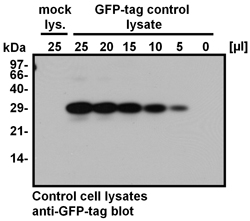
Western Blotting using the GFP-tag control-lysate and the mock-lysate.
HEK293 cells were transiently transfected either
with the empty expression vector (mock-lysate;
mock-lys.) or with an expression vector
encompassing the cDNA of green fluorescent
protein (GFP-tag control-lysate). 25 µl of the
mock-lysate and the indicated amounts of the
GFP-tag control-lysate (25, 20, 15, 10, 5, or 0 µl)
were separated by SDS-PAGE using a 15%
polyacrylamide-gel and then transfered onto a
PVDF membrane. The membrane was probed
with the polyclonal anti-GFP-tag antibody from tag-tools (dilution 1:500).
The GFP-tag bound primary antibody was
detected by incubation with HRP-coupled
protein A (dilution 1:10,000) and visualized
using chemiluminescence.
The X-ray film was exposed for 30 seconds.
Whereas a specific ~28 kDa band is detected
in the GFP-tag control-lysate at all volumes
(5 - 25 µl), no binding of the anti-GFP-tag
antibody to cellular proteins can be detected
in the lane, where 25 µl of the mock-lysate
was applied.
Use
The GFP-tag control-lysate and the mock-lysate
are for quality control in research applications only.
Not for diagnostic or therapeutic purpose.
![]()