navigation: home > Antikörper > Anti-GFP-tag
Anti-GFP-tag
Description
The GFP-tag, encoding the 26 kDa green fluorescent protein from the jellyfish Aequorea victoria, has revolutionized protein localization and live-cell imaging studies. However, in any experimental setup, expression of the correct sized GFP-fusion protein has to be verified by Western blotting. Our affinity-purified polyclonal antibody is ideally suited to detect GFP in Western Blotting applications. It can also be used to detect GFP-derived fluorescent proteins such as CFP, Cerulean, CyPet, YFP, or YPet. Furthermore, this polyclonal antibody has been shown to be superior to monoclonal antibodies, when it comes to immunoprecipitation of GFP.
Product
Immunogen:
Recombinant, full-length GFP derived from A. victoria
Antibody-type:
rabbit polyclonal
Concentration:
500 µg/ml
Concentration::
1 mg/ml
Purification:
Affinity purification by chromatography
on GFP-sepharose
Supplied buffer:
PBS, pH 7.2, containing 50% glycerol.
Shipping and storage
Shipping:
antibody is shipped in cold case
Storage:
antibody is stable for 1 month at 4°C. For prolonged storage, the antibody should be stored at -20°C. Aliquot to avoid repeated freeze-thaw-cycles. At -20°C, the product is stable for at least 1 year from shipment.
Use
For research use only. Not for diagnostic or therapeutic purpose.
Applications
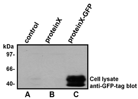
Western Blotting with the GFP-tag antibody
Lysates from human HEK293 cells transiently
transfected with the empty vector (control; lane A)
or a eukaryotic expression vector encoding
a ~50 kDa protein either without a tag
(proteinX; lane B) or with a carboxy-terminal
GFP-tag (proteinX-GFP; lane C) were
separated by SDS-PAGE and transfered onto
a PVDF membrane.
The membrane was probed with the anti-GFP-tag
antibody (1:500 dilution). Bound antibody was
visualized using horseradish peroxidase-coupled
Protein A (1:5,000 dilution) and chemiluminescence
detection. Exposure time 30 seconds. Each lane
contained equal amount of total protein.
Western Blotting: 1:500 – 1:2,000
Immunoprecipitation: 2 µg/sample
Optimal dilutions are dependent on conditions and should be determined by the user.
![]()