navigation: home > Antikörper > Anti-HA-tag
Anti-HA-tag
Description
The HA-tag is derived from a nona-peptide
within the influenza virus hemaglutinin protein.
The sequence of the nine amino acids
comprising the HA-tag (YPYDVPDYA) is not
present in any human or murine protein.
Therefore, the monoclonal antibody directed
against the HA-tag shows low background
binding to other proteins even in high
complexity samples such as whole
cell lysates.
The versatility of our monoclonal antibody
in Western Blotting, immunoprecipitation,
or immunofluorescence staining and the
inclusion of the HA-tag coding sequence in
many commercial expression vectors make
this antibody a must in every laboratory.
Product
Immunogen:
Synthetic peptide (YPYDVPDYA) derived
from the human influenza hemaglutinin protein
Antibody-type:
mouse monoclonal (clone TT7)
Isotype:
IgG2b
Concentration:
1 mg/ml
Purification:
Protein G affinity chromatography
Supplied buffer:
PBS, pH 7.2, containing 50% glycerol.
Shipping and storage
Shipping:
Antibody is shipped in cold case
Storage:
Antibody is stable for 1 month at 4°C. For prolonged storage, the antibody should be stored at -20°C. Aliquot to avoid repeated freezing and thawing. At -20°C, the product is stable for at least 1 year from shipment.
Use
For research use only. Not for diagnostic or therapeutic purpose.
Applications
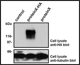
Western Blotting with the HA-tag antibody
Lysates from human HEK293 cells transiently
transfected with the empty vector (control; lane A)
or a eukaryotic expression vector encoding
a ~150 kDa protein either with a carboxy-terminal
HA-tag (proteinX-HA; lane B) or without a tag
(proteinX; lane C) were separated by SDS-PAGE
and transfered onto a PVDF membrane.
The membrane was probed with the anti-HA-tag
antibody (clone TT7; 1:2,000 dilution).
Bound antibody was visualized using horseradish
peroxidase-coupled Protein G (1:10,000 dilution)
and chemiluminescence detection.
Exposure time 30 seconds (upper panel).
The same samples were probed with an
anti-tubulin antibody to demonstrate equal loading
of the lysates (lower panel).
Western Blotting: 1:500 – 1:2,000
Immunoprecipitation: 2 µg/sample
Immunofluorescence staining: 1:200
(paraformaldehyde-fixed cells and tissues)
Immunohistochemistry: 1:100
(cryosections)
Optimal dilutions are dependent on experimental conditions and should be determined by the user.
![]()