navigation: home > Antikörper > Anti-phospho-tyrosine
Anti-phospho-tyrosine (Anti-pTyr) Neu!
Description
Phosphorylation of tyrosine residues within proteins is a post-translational modification involved in the regulation of almost every cellular process in eukaryotes. This reversible modification is brought about by one of the ~90 protein tyrosine kinases (PTKs) encoded in the mammalian genome, and is counterregulated by a similar complement of protein tyrosine phosphatases (PTPs). Our monoclonal antibody raised against a mixture of phospho-tyrosine (pTyr)-containing peptides has been proven to be useful for Western Blotting, immunostaining, and immunoprecipitation, making it one of the most versatile and specific reagents to monitor tyrosine phosphorylation of cellular proteins. Therefore, this antibody is an essential tool in every laboratory interested in signal transduction.
Product
Immunogen:
Synthetic peptides containing pTyr-residues in diverse sequence contexts.
Antibody-type:
mouse monoclonal (clone TT11)
Isotype:
IgG1
Concentration:
1 mg/ml
Purification:
Protein G affinity chromatography
Supplied buffer:
PBS, pH 7.2, containing 20% glycerol.
Shipping and storage
Shipping:
Antibody is shipped in cold case
Storage:
Antibody is stable for 1 month at 4°C.
For prolonged storage, the antibody
should be stored at -20°C.
Aliquot to avoid repeated freeze-thaw-cycles.
At -20°C, the product is stable for at least
1 year from shipment.
Use
For research use only. Not for diagnostic or therapeutic purpose.
Applications
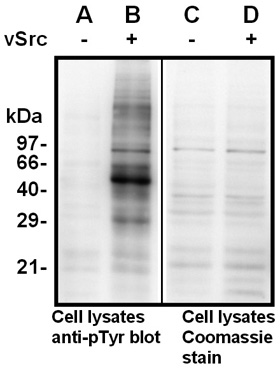
Western Blotting with the pTyr antibody
Lysates from human HEK293 cells transiently
transfected with the empty vector (control; lane A)
or a eukaryotic expression vector encoding
the oncogenic PTK v-Src (lane B) were separated
by SDS-PAGE and transfered onto a PVDF membrane.
The membrane was probed with pTyr antibody
(clone TT11; 1:1,000 dilution). Bound antibody was
visualized using horseradish peroxidase-coupled
Protein G (1:10,000 dilution) and chemiluminescence
detection. Exposure time 30 seconds.
The same lysates were loaded onto a separate gele
and proteins in the lysate were stained by Coomassie
Blue demonstrating equal amounts of total protein
in the sample without (lane C) and with v-Src (lane D).
Western Blotting: 1:500 – 1:2,000
Immunoprecipitation: 2 µg/sample
Immunofluorescence staining: 1:200
(paraformaldehyde-fixed cells and tissues)
Immunohistochemistry: 1:100
(cryosections)
Optimal dilutions are dependent on experimental conditions and should be determined by the user.
Anti-phospho-Tyrosin Antikörper![]()
![]()