navigation: home > control-lysates > phospho-tyrosine control-lysate
phospho-tyrosine control-lysate new!
Description
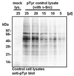
The phospho-tyrosine (pTyr) control-lysate was generated from transfected HEK293 cells, expressing the viral protein-tyrosine kinase v-Src. This virus-derived kinase is constitutively active and phosphorylates several cellular proteins, even in serum-starved cells. In contrast, tyrosine phosphorylation of cellular proteins in cells transfected with the empty vector (mock lysate) is barely detectable under these conditions. The cell-lysates were prepared using a lysis buffer, which contains multiple phosphatase- inhibitors to preserve the protein-tyrosine phosphory- lation in the cell lysates. In lysates from v-Src expressing cells (pTyr control lysate) cellular proteins with an apparent molecular weight of ~30, 55, and 75 kDa are the most intense bands detectable by Western Blotting with the anti-pTyr antibody.
Product
Whole cell lysate:
50 µl
Cell line:
HEK293 cells
Overexpressed protein:
v-Src , functioning as a constitutively-
active protein-tyrosine kinase
MW:
multiple pTyr proteins (~30, 55, and 75 kDa)
Lysis buffer:
60 mM Tris/HCL pH 6.8, 2% SDS,
5% mercaptoethanol,
50 µg/ml bromophenolblue, 10% glycerine
Application:
For SDS-PAGE/Western Blotting
20 µl of the lysates should be loaded per
lane of a 10% polyacrylamide-gel.
Shipping and storage
Shipping:
The lysate is shipped in cold case.
Storage:
Lysate is stable for 1 week at 4°C.
For prolonged storage, the lysate should be
stored at -80°C. Aliquot to avoid repeated freezing
and thawing.
At -80°C, the product is stable for at least 1 year
from shipment.
Application in quality control

Western Blotting using the pTyr control lysate
and the mock lysate.
HEK293 cells were transiently transfected either
with a eukaryotic expression vector, encoding the
cDNA for the protein tyrosine kinase v-Src (pTyr
control lysate), or with the empty expression
vector (mock lysate; mock lys.).
The indicated amounts of the pTyr control-lysate
(25, 20, 15, 10, or 5 µl) or 25 µl of the mock lysate,
respectively, were separated by SDS-PAGE on a
10% polyacrylamide-gel and then transfered onto
a PVDF membrane. The membrane was probed
with the monoclonal anti-pTyr antibody from tag-tools (clone TT11; dilution 1:1.000).
The bound antibody was detected by incubation
with HRP-coupled protein G (dilution 1:10.000) and
visualized using chemiluminescence. The X-ray
film was exposed for 15 seconds.
Multiple tyrosine phosphorylated proteins are
visible in 5 - 25 µl of the lysates from v-Src expressing
cells (pTyr control lysate), with the most prominent
bands at ~30, 55, and 75 kDa. In contrast, barely any
tyrosine phosphorylation is detectable in 25 µl of
the mock lysate derived from control transfected,
serum-starved cells.
Use
The pTyr control lysate and the mock lysate
are for quality control in research applications only.
They are not intended for diagnostic or therapeutic
purposes.
Anti-phospho-tyrosine antibody
![]()

